
Bioactive Compound from Nypa fruticans Endophytic Fungi: Isolation, Identification, Biological Screening, and In Silico Studies
DOI:
https://doi.org/10.11113/mjfas.v22n5.5838Keywords:
Nypa fruticans, endophytic fungi, Antibacterial, Antioxidant, molecular dockingAbstract
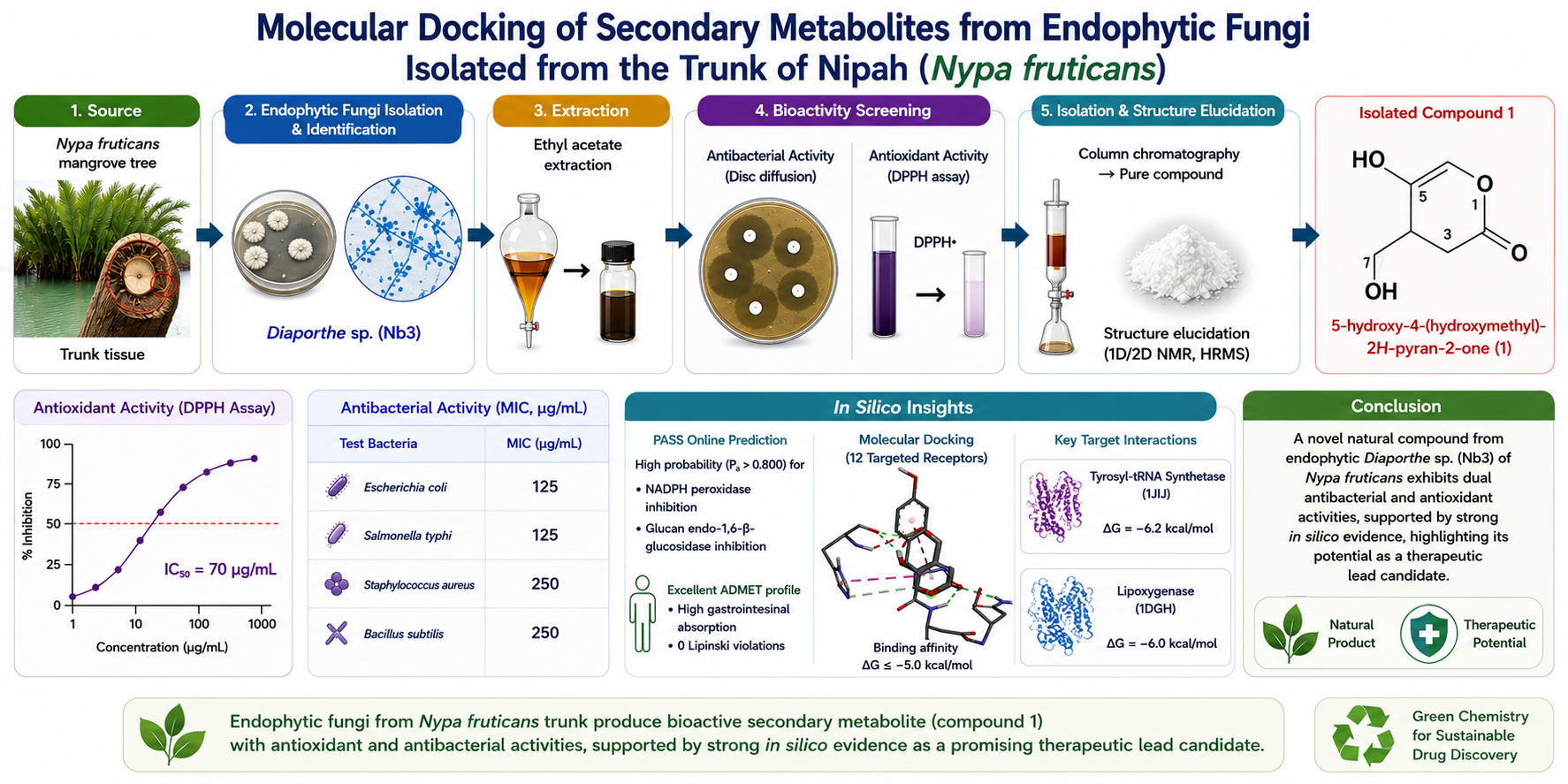
Nypa fruticans is one of the unique and dominant mangrove species of tropical coastal areas that is traditionally used for the treatment of many different illnesses. The objective of this study was to isolate and identify bioactive secondary metabolite from endophytic fungi from trunk of Nypa fruticans. The investigation included the isolation and morphological identification of endophytic fungi, followed by ethyl acetate extraction. The crude extracts were screened for antibacterial activity by disc diffusion method and antioxidant potential by DPPH assay. Purification of the highly active Diaporthe sp. (Nb3) extract by column chromatography yielded a pure crystalline compound, which was elucidated by 1D/2D NMR and mass spectrometry as 5-hydroxy-4-(hydroxymethyl)-2H-pyran-2-one (compound 1). Compound 1 showed a significant antioxidant scavenging activity under our assay conditions (IC50 = 70 μg/mL) and moderate antibacterial activity against Escherichia coli (MIC 125 μg/mL), Salmonella typhi (MIC 125 μg/mL), Staphylococcus aureus (MIC 250 μg/mL) and Bacillus subtilis (MIC 250 μg/mL). To rationalize these biological networks, comprehensive in silico screens were performed. PASS Online profiling predicted high probabilities (Pa>0.800) for NADPH peroxidase and glucan endo-1,6-beta-glucosidase inhibition with an excellent ADMET profile characterized by high gastrointestinal absorption and zero Lipinski violations. Molecular docking analysis also showed spontaneous binding configurations (ΔG≤-5.0 kcal/mol) on 12 targeted receptors. These dual mechanisms were stabilized by strong binding affinity toward the antibacterial target tyrosyl-tRNA synthetase (PDB ID: 1JIJ, ΔG=−6.2 kcal/mol) and key cytoprotective regulators such as lipoxygenase (PDB ID: 1DGH, ΔG=−6.0 kcal/mol). The computational alignment ultimately confirms the isolated compound as a potent therapeutic lead candidate.
References
[1] S. K. Ahmed et al., “Antimicrobial resistance: Impacts, challenges, and future prospects,” J. Med. Surgery, Public Heal., vol. 2, no. March, p. 100081, 2024, doi: 10.1016/j.glmedi.2024.100081.
[2] T. R. Walsh, A. C. Gales, R. Laxminarayan, and P. C. Dodd, “Antimicrobial Resistance: Addressing a Global Threat to Humanity,” PLoS Med., vol. 20, no. 7 July, pp. 12–15, 2023, doi: 10.1371/journal.pmed.1004264.
[3] I. Frost, A. Balachandran, S. Paulin-Deschenaux, H. Sati, and M. Hasso-Agopsowicz, “The approach of World Health Organization to articulate the role and assure impact of vaccines against antimicrobial resistance,” Hum. Vaccines Immunother., vol. 18, no. 6, pp. 0–4, 2022, doi: 10.1080/21645515.2022.2145069.
[4] C. S. Ho et al., “Antimicrobial resistance: a concise update,” The Lancet Microbe, vol. 6, no. 1, pp. 1–14, 2025, doi: 10.1016/j.lanmic.2024.07.010.
[5] T. B. Tufa, F. Regassa, K. Amenu, J. A. Stegeman, and H. Hogeveen, “Livestock producers’ knowledge, attitude, and behavior (KAB) regarding antimicrobial use in Ethiopia,” Front. Vet. Sci., vol. 10, pp. 1–20, 2023, doi: 10.3389/fvets.2023.1167847.
[6] C. E. Flynn and J. Guarner, “Emerging Antimicrobial Resistance,” Mod. Pathol., vol. 36, no. 9, p. 100249, 2023, doi: 10.1016/j.modpat.2023.100249.
[7] F. O. Ohiagu, P. C. Chikezie, T. O. Maduka, C. M. Chikezie, O. Nwaiwu, and K. R. Paudel, “Antioxidants, Radical Scavengers, and Their Impact on Oxidative Stress,” Free Radicals Antioxidants, vol. 14, no. 2, pp. 62–85, 2025, doi: 10.5530/fra.2024.2.7.
[8] İ. Gulcin, Antioxidants: a comprehensive review, vol. 99, no. 5. 2025. doi: 10.1007/s00204-025-03997-2.
[9] S. T. Omaye, “Antioxidants: Looking forward after a decade,” Antioxidants, vol. 10, no. 12, 2021, doi: 10.3390/antiox10121992.
[10] J. Flieger, W. Flieger, and J. Baj, “Antioxidants : Classification , Natural Sources , Activity / Capacity,” Materials (Basel)., vol. 14, no. 4135, pp. 1–54, 2021, [Online]. Available: https://www.mdpi.com/journal/materials
[11] M. E. Rusu, I. Fizeșan, L. Vlase, and D. S. Popa, “Antioxidants in Age-Related Diseases and Anti-Aging Strategies,” Antioxidants, vol. 11, no. 10, pp. 1–5, 2022, doi: 10.3390/antiox11101868.
[12] B. Alam et al., “Endophytic Fungi: From Symbiosis to Secondary Metabolite Communications or Vice Versa?,” Front. Plant Sci., vol. 12, no. December, pp. 1–24, 2021, doi: 10.3389/fpls.2021.791033.
[13] Q. xi Li, F. cheng Lin, and Z. zhu Su, “Endophytic fungi-Big player in plant-microbe symbiosis,” Curr. Plant Biol., vol. 42, no. April, p. 100481, 2025, doi: 10.1016/j.cpb.2025.100481.
[14] J. Wen et al., “Endophytic Fungi: An Effective Alternative Source of Plant‐Derived Bioactive Compounds for Pharmacological Studies,” J. Fungi, vol. 8, no. 2, 2022, doi: 10.3390/jof8020205.
[15] C. Liao et al., Challenges and update on fungal endophytes: classification, definition, diversity, ecology, evolution and functions, vol. 131, no. 1. Springer Netherlands, 2025. doi: 10.1007/s13225-025-00550-5.
[16] M. Zhang, C. Zhong, X. Lv, Z. Fang, C. Cheng, and J. Hao, “Germplasm Resource Status and Seed Adaptability of Nypa fruticans Wurmb, an Endangered Species in China,” Forests, vol. 15, no. 8, pp. 1–16, 2024, doi: 10.3390/f15081396.
[17] G. D. Nugroho et al., “Review: Phytochemical composition, medicinal uses and other utilization of Nypa fruticans,” Int. J. Bonorowo Wetl., vol. 10, no. 1, pp. 51–65, 2022, doi: 10.13057/bonorowo/w100105.
[18] S. S. A. Nasution, Elfita, H. Widjajanti, and Ferlinahayati, “Diversity, bioactivity, and phytochemistry of endophytic fungi in various organs of nipa palm (Nypa fruticans) mangrove,” Biodiversitas, vol. 25, no. 10, pp. 3928–3942, 2024, doi: 10.13057/biodiv/d251053.
[19] A. Loilong, J. Sakayaroj, N. Rungjindamai, R. Choeyklin, and E. B. G. Jones, “Biodiversity of fungi on the palm Nypa fruticans,” Mar. Fungi Fungal-like Org., pp. 273–290, 2012, doi: 10.1515/9783110264067.273.
[20] P. Toppo, P. Jangir, N. Mehra, R. Kapoor, and P. Mathur, “Bioprospecting of endophytic fungi from medicinal plant Anisomeles indica L. for their diverse role in agricultural and industrial sectors,” Sci. Rep., vol. 14, no. 1, pp. 1–18, 2024, doi: 10.1038/s41598-023-51057-5.
[21] N. H. Caicedo, A. F. Davalos, P. A. Puente, A. Y. Rodríguez, and P. A. Caicedo, “Antioxidant activity of exo-metabolites produced by Fusarium oxysporum: An endophytic fungus isolated from leaves of Otoba gracilipes,” Microbiologyopen, vol. 8, no. 10, pp. 1–7, 2019, doi: 10.1002/mbo3.903.
[22] E. Charria-Girón et al., “Evaluation of the Antibacterial Activity of Crude Extracts Obtained From Cultivation of Native Endophytic Fungi Belonging to a Tropical Montane Rainforest in Colombia,” Front. Microbiol., vol. 12, no. September, 2021, doi: 10.3389/fmicb.2021.716523.
[23] T. Watanabe, Pictorial Atlas of Soil and Seed Fungi. 2010. doi: 10.1201/ebk1439804193.
[24] D. H. Larone, L. F. Westblade, E. M. Burd, S. R. Lockhart, G. W. Procop, and T. J. Walsh, Larone’s medically important fungi: A guide to identification (7th ed.). John Wiley & Sons, 2023. doi: 10.1128/9781555819880.
[25] L. LaPierre, “Antimicrobial Susceptibility Testing Laboratory Guide : Methodologies for,” APEC Proj. CTI 24 2017A, no. May, pp. 1–19, 2020.
[26] S. Baliyan et al., “Determination of Antioxidants by DPPH Radical Scavenging Activity and Quantitative Phytochemical Analysis of Ficus religiosa,” Molecules, vol. 27, no. 4, 2022, doi: 10.3390/molecules27041326.
[27] S. Dallakyan and A. J. Olson, “Small-Molecule Library Screening by Docking with PyRx,” vol. 1263, pp. 243–250, 2015, doi: 10.1007/978-1-4939-2269-7.
[28] L. Schrödinger, “The PyMOL Molecular Graphics System, Version 3.0,” 2023.
[29] D. S. BIOVIA, “Discovery Studio Modeling Environment,” 2025, Dassault Systèmes, San Diego.
[30] B. Kowalska-Krochmal and R. Dudek-Wicher, “The minimum inhibitory concentration of antibiotics: Methods, interpretation, clinical relevance,” Pathogens, vol. 10, no. 2, pp. 1–21, 2021, doi: 10.3390/pathogens10020165.
[31] Elfita, R. Oktiansyah, Mardiyanto, H. Widjajanti, A. Setiawan, and S. S. A. Nasution, “Bioactive Compounds of Endophytic Fungi Lasiodiplodia theobromae Isolated From The Leaves of Sungkai (Peronema canescens),” Biointerface Res. Appl. Chem., vol. 13, no. 6, Dec. 2023, doi: 10.33263/BRIAC136.530.
[32] A. Lin, X. Lu, Y. Fang, T. Zhu, Q. Gu, and W. Zhu, “NOTE Two New 5-Hydroxy-2-pyrone Derivatives Isolated from a Marine-derived Fungus Aspergillus flavus,” vol. 61, no. 4, pp. 245–249, 2008.
[33] A. N. M. Gubran and M. Ali, “Narrative review : Cell wall synthesis inhibitors with an emphasis on mode of actions , resistance mechanisms , and clinical utility and dosages,” The Microbe, vol. 11, no. March, p. 100700, 2026, doi: 10.1016/j.microb.2026.100700.
[34] A. Yadav and R. K. Salar, “Next Nanotechnology Tyrosinase inhibitors : Molecular mechanisms , therapeutic potentials , and translational applications in dermatology and beyond,” Next Nanotechnol., vol. 9, no. February, p. 100452, 2026, doi: 10.1016/j.nxnano.2026.100452.
[35] X. Qiu et al., “Crystal structure of Staphylococcus aureus tyrosyl-tRNA synthetase in complex with a class of potent and specific inhibitors,” no. 2001, pp. 2008–2016, 2016, doi: 10.1101/ps.18001.).
[36] N. Schormann et al., “The catalytic domains of Streptococcus mutans glucosyltransferases : a structural analysis research communications,” pp. 119–127, 2023, doi: 10.1107/S2053230X23003199.
[37] X. Li et al., “Bioorganic & Medicinal Chemistry Letters Structure-based design of new DHFR-based antibacterial agents :,” Bioorg. Med. Chem. Lett., vol. 21, no. 18, pp. 5171–5176, 2011, doi: 10.1016/j.bmcl.2011.07.059.
[38] W. Deng and C. L. M. J. V. Verlinde, “Evaluation of Different Virtual Screening Programs for Docking in a Charged Binding Pocket,” J. Chem. Inf. Model., vol. 48, no. 10, pp. 2010–2020, 2008, doi: 10.1021/ci800154w.Evaluation.
[39] N. Bernardo-garcia et al., “Allostery , Recognition of Nascent Peptidoglycan and Crosslinking of the Cell Wall by the Essential Penicillin- Binding Protein 2x of Streptococcus pneumoniae Allostery , Recognition of Nascent Peptidoglycan and Crosslinking of the Cell Wall by the Essent,” ACS Chem. Biol., 2018, doi: 10.1021/acschembio.7b00817.
[40] P. Panchaud et al., “Discovery and Optimization of Isoquinoline Ethyl Ureas as Antibacterial Agents,” J. Med. Chem., 2017, doi: 10.1021/acs.jmedchem.6b01834.
[41] C. . Kavanagh, K.L.; Johansson, C.; Papagrigoriou, E.; Kochan, G.; Umeano, M. . Gileadi, O.; von Delft, F.; Weigelt, J.; Arrowsmith, C.H.; Sundstrom, and U. . S. G. C. (SGC) Edwards, A.; Oppermann, “Crystal structure of human glutathione peroxidase 7,” 2007.
[42] C. Aguirre, T. ten Brink, J.-F. Guichou, O. Cala, and I. Krimm, “Comparing Binding Modes of Analogous Fragments Using NMR in Fragment-Based Drug Design : Application to PRDX5,” vol. 9, no. 7, pp. 1–11, 2014, doi: 10.1371/journal.pone.0102300.
[43] C. D. Putnam, A. S. Arvai, Y. Bourne, J. A. Tainer, and J. Aiguier, “Active and Inhibited Human Catalase Structures : Ligand and NADPH Binding and Catalytic Mechanism,” 2000.
[44] S. N. Savvides and P. A. Karplus, “Kinetics and Crystallographic Analysis of Human Glutathione Reductase in Complex with a Xanthene Inhibitor *,” J. Biol. Chem., vol. 271, no. 14, pp. 8101–8107, 1996, doi: 10.1074/jbc.271.14.8101.
[45] H. tae Kim, H. Cha, and K. Y. Hwang, “Structural insight into the inhibition of carbonic anhydrase by the COX-2-selective inhibitor polmacoxib ( CG100649 ),” Biochem. Biophys. Res. Commun., vol. 478, no. 1, pp. 1–6, 2016, doi: 10.1016/j.bbrc.2016.07.114.
[46] T. Saito et al., “p62/Sqstm1 promotes malignancy of HCV-positive hepatocellular carcinoma through Nrf2-dependent metabolic reprogramming,” Nat. Commun., 2016, doi: 10.1038/ncomms12030.
Downloads
Published
Issue
Section
License
Copyright (c) 2026 Sakinah Salman Ahmad Nasution, Elfita, Hary Widjajanti, Ferlinahayati, Sofa Fajriah

This work is licensed under a Creative Commons Attribution-NonCommercial 4.0 International License.