
Multi-omics profiling Profiling nominates Nominates caspaseCaspase-6 (CASP6) as a clinically actionablecCandidate prognostic Prognostic signaturebBiomarker in kidney Kidney renal Renal clear Clear cell Cell carcinomaCarcinoma
DOI:
https://doi.org/10.11113/mjfas.v22n6.4691Keywords:
caspase, anticancer, prognostic potential, CASP6, kidney renal clear cell carcinomaAbstract
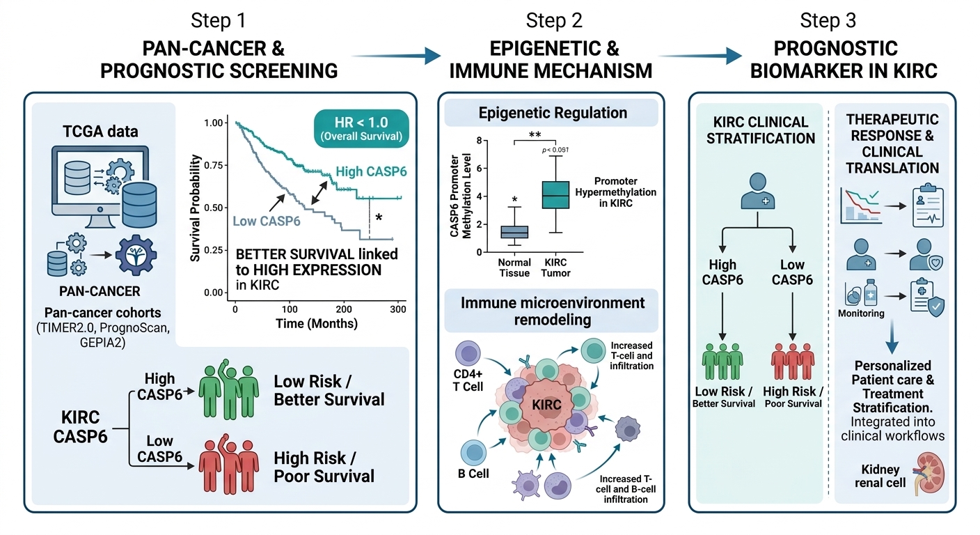
Caspase (CASP) family members have been proposedare frequently employed as pharmacodynamic biomarkers for monitoringto evaluate anticancer therapeutic responses, yet their prognostic relevance across malignancies remains incompletely characterizedunderexplored. The present investigation systematically assessed the prognostic potentialclinical utility of seven proapoptosis-associated caspase genes (CASP2/3/6/7/8/9/10) across pan-cancer cohorts usingas pan-cancer prognostic indicators. Iintegrative analysis of TIMER2.0, PrognoScan, and GEPIA2 databases. The screening identified CASP6 and CASP9 as candidate survival-associated geneskey survival predictors:; elevated CASP6 expression correlated with favorable outcomes in kidney renal clear cell carcinoma (KIRC) and colon adenocarcinoma, while high CASP9 levels were associated with improvedpredicted ameliorative survival in kidney renal papillary cell carcinoma (KIRP). TIMER2.0 immune events analysis revealed CASP6-associated distinct immune microenvironment remodeling in KIRC, characterized by coordinated infiltration withof B cells, CD4+ T cells, and myeloid populations. UALCAN methylation profiling demonstrated tumor-specific promoter hypermethylation of CASP6 in KIRC and CASP9 in KIRP, which is strongly correlated with transcriptional downregulation. cBioPortal analysis identified missense mutations (CASP6- E16G/K238R in colon adenocarcinoma; CASP9- S133F in KIRP) classified as variants of uncertain significance. Through multi-omics integrationvalidation, CASP6 emerged as a candidaterobust transcriptional biomarker forin KIRC, with expression levels stratifyingshowing potential associations with therapeutic response probabilities. These findings establishedsuggest that caspase expression signature may serve as complementary tools for prognostic stratification in renal malignancies.
References
[1] A. Boice, L. Bouchier-Hayes, Biochim. Biophys. Acta Mol. Cell Res. 1867 (2020) 118688.
[2] L. Dorstyn, S. Kumar, Methods Mol. Biol. 1133 (2014) 71–87.
[3] A.N. Brown-Suedel, L. Bouchier-Hayes, Front. Cell Dev. Biol. 8 (2020) 610022.
[4] M. Asadi, S. Taghizadeh, E. Kaviani, O. Vakili, M. Taheri-Anganeh, M. Tahamtan, A. Savardashtaki, Biotechnol. Appl. Biochem. 69 (2022) 1633–1645.
[5] A.A. Bhat, R. Thapa, O. Afzal, N. Agrawal, W.H. Almalki, I. Kazmi, S.I. Alzarea, A.S.A. Altamimi, et al., Int. J. Biol. Macromol. 242 (2023) 124832.
[6] M. Zheng, R. Karki, P. Vogel, T.D. Kanneganti, Cell 181 (2020) 674–687.
[7] K. Nozaki, V.I. Maltez, M. Rayamajhi, A.L. Tubbs, J.E. Mitchell, C.A. Lacey, C.K. Harvest, L. Li, et al., Nature 606 (2022) 960–967.
[8] M. Fritsch, S.D. Günther, R. Schwarzer, M.C. Albert, F. Schorn, J.P. Werthenbach, L.M. Schiffmann, N. Stair, et al., Nature 575 (2019) 683–687.
[9] A. Unnisa, N.H. Greig, M.A. Kamal, Curr. Neuropharmacol. 21 (2023) 1001–1012.
[10] B. Kim, S.K. Srivastava, S.H. Kim, Expert Opin. Ther. Targets 19 (2015) 113–127.
[11] J. Yuan, Y. Deng, Y. Zhang, X. Gan, S. Gao, H. Hu, S. Hu, J. Hu, et al., Anim. Reprod. Sci. 200 (2019) 86–95.
[12] A. Mohr, L. Deedigan, S. Jencz, Y. Mehrabadi, L. Houlden, S.M. Albarenque, R.M. Zwacka, Cell Death Differ. 25 (2018) 340–352.
[13] C. Jiao, W. Chen, X. Tan, H. Liang, J. Li, H. Yun, C. He, J. Chen, et al., J. Ethnopharmacol. 247 (2020) 112256.
[14] N.H. Kim, M.J. Xin, J.Y. Cha, S.J. Ji, S.U. Kwon, H.K. Jee, M.R. Park, Y.S. Park, et al., Am. J. Chin. Med. 45 (2017) 319–335.
[15] A. Mohammadi, B. Mansoori, P.C. Baradaran, S.C. Baradaran, B. Baradaran, J. Gastrointest. Cancer 48 (2017) 333–340.
[16] H. Ke, X. Wang, Z. Zhou, W. Ai, Z. Wu, Y. Zhang, J. Ethnopharmacol. 264 (2021) 113363.
[17] M.I. Alhazmi, T.N. Hasan, G. Shafi, A.H. Al-Assaf, M.A. Alfawaz, A.A. Alshatwi, Asian Pac. J. Cancer Prev. 15 (2014) 9655–9660.
[18] A.A. Al-Fatlawi, A.A. Al-Fatlawi, M. Irshad, Rahisuddin, A. Ahmad, Pharm. Biol. 53 (2015) 104–109.
[19] W. Lan, S. Wan, W. Gu, H. Wang, S. Zhou, Cell Biochem. Biophys. 70 (2014) 1459–1467.
[20] B.B. Aggarwal, A. Kumar, A.C. Bharti, Anticancer Res. 23 (2003) 363–398.
[21] G.M. Calaf, D. Roy, Int. J. Oncol. 51 (2017) 1590–1600.
[22] P.E. Czabotar, A.J. Garcia-Saez, Nat. Rev. Mol. Cell Biol. 24 (2023) 732–748.
[23] J. Kale, E.J. Osterlund, D.W. Andrews, Cell Death Differ. 25 (2018) 65–80.
[24] M. Olsson, B. Zhivotovsky, Cell Death Differ. 18 (2011) 1441–1449.
[25] G. Yu, H. Chi, G. Zhao, Y. Wang, Front. Oncol. 14 (2024) 1375334.
[26] T. Li, J. Fu, Z. Zeng, D. Cohen, J. Li, Q. Chen, B. Li, X.S. Liu, Nucleic Acids Res. 48 (2020) W509–W514.
[27] H. Mizuno, K. Kitada, K. Nakai, A. Sarai, BMC Med. Genomics 2 (2009) 18.
[28] Z. Tang, B. Kang, C. Li, T. Chen, Z. Zhang, Nucleic Acids Res. 47 (2019) W556–W560.
[29] J. Gao, B.A. Aksoy, U. Dogrusoz, G. Dresdner, B. Gross, S.O. Sumer, Y. Sun, A. Jacobsen, et al., Sci. Signaling 6 (2013) pl1.
[30] H.A. Shihab, J. Gough, D.N. Cooper, I.N. Day, T.R. Gaunt, Bioinformatics 29 (2013) 1504–1510.
[31] D.S. Chandrashekar, B. Bashel, S.A.H. Balasubramanya, C.J. Creighton, I. Ponce-Rodriguez, B. Chakravarthi, S. Varambally, Neoplasia 19 (2017) 649–658.
[32] S. Vasaikar, C. Huang, X. Wang, V.A. Petyuk, S.R. Savage, B. Wen, Y. Dou, Y. Zhang, et al., Cell 177 (2019) 1035–1049.
[33] G. Guo, Y. Gui, S. Gao, A. Tang, X. Hu, Y. Huang, W. Jia, Z. Li, et al., Nat. Genet. 44 (2012) 17–19.
[34] Y. Sato, T. Yoshizato, Y. Shiraishi, S. Maekawa, Y. Okuno, T. Kamura, T. Shimamura, A. Sato-Otsubo, et al., Nat. Genet. 45 (2013) 860–867.
[35] M. Gerlinger, S. Horswell, J. Larkin, A.J. Rowan, M.P. Salm, I. Varela, R. Fisher, N. McGranahan, et al., Nat. Genet. 46 (2014) 225–233.
[36] J.B. Pakish, Q. Zhang, Z. Chen, H. Liang, G.B. Chisholm, Y. Yuan, S.C. Mok, R.R. Broaddus, et al., Clin. Cancer Res. 23 (2017) 4473–4481.
[37] S. Zhang, E. Zhang, J. Long, Z. Hu, J. Peng, L. Liu, F. Tang, L. Li, et al., Cancer Sci. 110 (2019) 1564–1572.
[38] H. Gao, Y. Yao, W. Li, Z. Xu, W. Hu, K. Luo, P. Chen, W. Shang, et al., Adv. Sci. 12 (2025) e2411375.
[39] K. Liu, M. Wang, D. Li, N.T. Duc Duong, Y. Liu, J. Ma, K. Xin, Z. Zhou, Front. Immunol. 15 (2024) 1502855.
[40] L. Qi, L. Wang, M. Jin, M. Jiang, L. Li, Y. Li, Immunology 169 (2023) 245–259.
[41] Y. Lin, M. Sheng, H. Qin, P. Zhang, C. Wang, W. Fu, X. Meng, D. Wang, et al., Cell Commun. Signaling 21 (2023) 282.
[42] G.C. Hutterer, M. Pichler, T.F. Chromecki, K.A. Strini, T. Klatte, K. Pummer, M. Remzi, S. Mannweiler, et al., Histopathology 63 (2013) 309–315.
[43] X. Liu, S. Wu, Y. Yang, M. Zhao, G. Zhu, Z. Hou, Biomed. Pharmacother. 95 (2017) 55–61.
[44] G. Sokratous, S. Polyzoidis, K. Ashkan, Hum. Vaccines Immunother. 13 (2017) 2575–2582.
[45] P. Jia, Z. Zhao, Briefings Bioinf. 18 (2017) 413–425.
[46] J.W. Lee, M.R. Kim, Y.H. Soung, S.W. Nam, S.H. Kim, J.Y. Lee, N.J. Yoo, S.H. Lee, APMIS 114 (2006) 646–650.
[47] H. Heyn, M. Esteller, Nat. Rev. Genet. 13 (2012) 679–692.
[48] F. Arenas, I. Hervías, E. Sáez, S. Melero, J. Prieto, A. Parés, J.F. Medina, JHEP Rep. 1 (2019) 145–153.
[49] J. Wu, K. He, Y. Zhang, J. Song, Z. Shi, W. Chen, Y. Shao, Gene 687 (2019) 193–199.
[50] J. Hu, Y. Chen, L. Gao, C. Ge, X. Xie, P. Lei, Y. Zhang, P. Liang, Front. Genet. 13 (2022) 851384.
[51] S. Qian, H. Song, L. Huang, H. Hua, X. Zhang, Z. Li, M. Pu, H. Huang, et al., Cell. Mol. Biol. Lett. 30 (2025) 142.
[52] J. Coté, S. Dupuis, Z. Jiang, J.Y. Wu, Proc. Natl. Acad. Sci. U. S. A. 98 (2001) 938–943.
[53] N. Droin, M. Beauchemin, E. Solary, R. Bertrand, Cancer Res. 60 (2000) 7039–7047.
[54] P. Vito, T. Ghayur, L. D'Adamio, J. Biol. Chem. 272 (1997) 28315–28320.
[55] E. Devarajan, A.A. Sahin, J.S. Chen, R.R. Krishnamurthy, N. Aggarwal, A.M. Brun, A. Sapino, F. Zhang, et al., Oncogene 21 (2002) 8843–8851.
[56] B. Foveau, L. Van Der Kraak, N. Beauchemin, S. Albrecht, A.C. LeBlanc, PLoS One 9 (2014) e114270.
[57] Y. Krelin, L. Zhang, T.B. Kang, E. Appel, A. Kovalenko, D. Wallach, Cell Death Differ. 15 (2008) 1350–1355.
[58] M.P. Boldin, T.M. Goncharov, Y.V. Goltsev, D. Wallach, Cell 85 (1996) 803–815.
[59] D. Himeji, T. Horiuchi, H. Tsukamoto, K. Hayashi, T. Watanabe, M. Harada, Blood 99 (2002) 4070–4078.
[60] R.P. Graf, N. Keller, S. Barbero, D. Stupack, Curr. Mol. Med. 14 (2014) 246–254.
[61] W.S. Park, J.H. Lee, M.S. Shin, J.Y. Park, H.S. Kim, J.H. Lee, Y.S. Kim, S.N. Lee, et al., Oncogene 21 (2002) 2919–2925.
[62] Y. Shikama, M. Yamada, T. Miyashita, Eur. J. Immunol. 33 (2003) 1998–2006.
[63] Y. Shikama, L. Shen, M. Yonetani, J. Miyauchi, T. Miyashita, M. Yamada, Biochem. Biophys. Res. Commun. 291 (2002) 484–493.
[64] H. Wang, P. Wang, X. Sun, Y. Luo, X. Wang, D. Ma, J. Wu, Biochim. Biophys. Acta 1770 (2007) 1528–1537.
Downloads
Published
Issue
Section
License
Copyright (c) 2026 Shuai Ma, Tian Zhang, Yang Liu, Yueying Xu, Jianjun Cheng, Chunying Zhao, Yu Cao, Chunhan Qiao

This work is licensed under a Creative Commons Attribution-NonCommercial 4.0 International License.