
Anti-inflammatory, Antioxidant, and In Vitro Wound Healing Activities of Parkia speciosa Peel Extract with Application in Topical Formulation and Stability Assessment
DOI:
https://doi.org/10.11113/mjfas.v22n6.4689Keywords:
Parkia speciosa peel, nitric oxide inhibition, antioxidant activity, wound healing, topical formulationAbstract
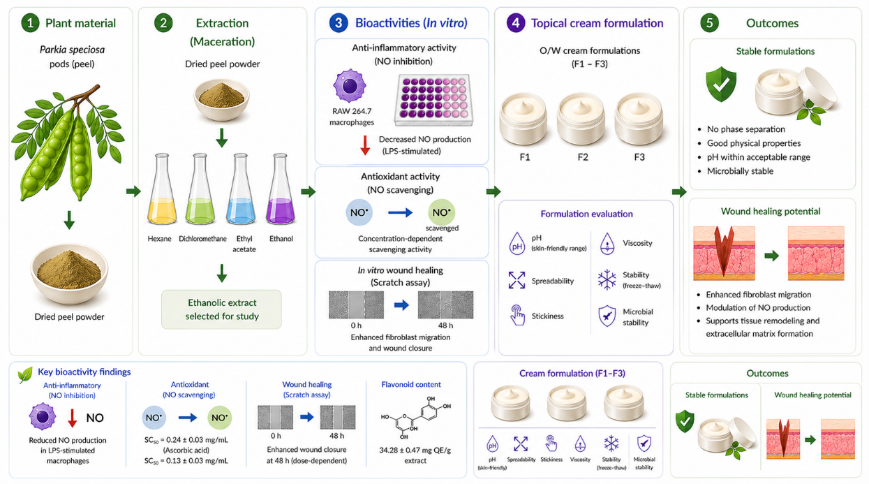
This study evaluated the anti-inflammatory, antioxidant, and in vitro wound healing activities of Parkia speciosa peel extract and its application in topical formulations. Peel powders were extracted separately using hexane, dichloromethane, ethyl acetate, and ethanol by maceration. Based on preliminary screening, the ethanol extract was selected for further analysis due to its higher bioactive content. Flavonoid content was determined using the aluminium chloride colorimetric method. Anti-inflammatory activity was assessed through nitric oxide (NO) inhibition in lipopolysaccharide (LPS)-stimulated RAW 264.7 macrophage cells, while antioxidant activity was evaluated using an NO scavenging assay. The extract exhibited a flavonoid content of 34.28 ± 0.47 mg QE/g extract and showed concentration-dependent NO scavenging activity with an SC₅₀ value of 0.24 ± 0.03 mg/mL, compared to 0.13 ± 0.03 mg/mL for ascorbic acid. The extract also significantly reduced NO production in LPS-stimulated macrophages. In vitro wound healing potential was evaluated using a human fibroblast scratch assay. Lower concentrations (7.81 and 15.63 µg/mL) enhanced wound closure at 48 h, whereas a higher concentration (31.25 µg/mL) promoted early cell migration. Three cream formulations (F1–F3) containing the extract were developed and evaluated for physical properties and stability. Formulation F3 exhibited improved spreadability, reduced stickiness, and maintained stability without phase separation after six freeze–thaw cycles, with pH remaining within the acceptable dermatological range. These findings indicate that P. speciosa peel extract possesses anti-inflammatory, antioxidant, and wound healing activities and can be incorporated into stable topical formulations. Further studies, including in vivo evaluation, are required to confirm its efficacy and safety.
References
[1] K. Yusoff, S.F.Z. Japar, H.J. Abdul, J. Kamsiah, Biomed. Pharmacother. 96 (2017) 291–298.
[2] N.H. Mustafa, A. Ugusman, J. Jalil, J. Appl. Pharm. Sci. 8 (2018) 152–158.
[3] J.S. Gui, J. Jalil, Z. Jubri, Cytotechnology 71 (2019) 79–89.
[4] K. Azemi, M.L. Nordin, K.A. Hambali, Antioxidants 11 (2022) 431.
[5] P. Krzyszczyk, R. Schloss, A. Palmer, F. Berthiaume, Front. Physiol. 9 (2018) 419.
[6] A. Joorabloo, T. Liu, J. Nanobiotechnol. 20 (2022) 407.
[7] S.Y. Kim, M.G. Nair, Immunol. Cell Biol. 97 (2019) 450–460.
[8] A. Hassanshahi, M. Moradzad, S. Ghalamkari, Cells 11 (2022) 2953.
[9] L. Sautebin, Fitoterapia 71 (2000) 48–57.
[10] N. Muhamad, P.S. Sinchai, U. Tansom, J. Adv. Dev. Eng. Sci. 14 (2024) 101–110.
[11] C. Chang, M.Yang, H. Wen, J. Chern, J. Food Drug Anal. 10 (2002) 178–182.
[12] P. Griess, Ann. Chem. Pharm. 106 (1858) 123–125.
[13] L. Marcocci, J.J. Maguire, M.T. Droy-Lefaix, L. Packer, Biochem. Biophys. Res. Commun. 201 (1994) 748–755.
[14] G. Cory, in: C. Wells, M. Parsons (Eds.), Cell Migration, Methods Mol. Biol. 769, Humana Press, Totowa, 2011, p. 25–30.
[15] A.O. Barel, M. Paye, H.I. Maibach, Handbook of Cosmetic Science and Technology, 4th ed., CRC Press, Boca Raton, 2014.
[16] M.E. Aulton, K.M.G. Taylor, Aulton’s Pharmaceutics: The Design and Manufacture of Medicines, 5th ed., Elsevier, Edinburgh, 2018.
[17] W. Siegert, Cosmetics 6 (2019) 25.
[18] S.M. Jafari, Adv. Colloid Interface Sci. 254 (2018) 1–17.
[19] M.S.M. Saleh, Int. J. Mol. Sci. 22 (2021) 618.
[20] F.H. Builders, Phytomedicine 20 (2021) 521–530.
[21] S. Guo, L.A. DiPietro, J. Dent. Res. 89 (2010) 219–229.
[22] H.A. Wallace, B.M. Basehore, P.M. Zito, Wound Healing Phases, StatPearls Publishing, Treasure Island, 2023.
[23] J. Dai, R.J. Mumper, Molecules 15 (2010) 7313–7352.
[24] J.S. Beckman, W.H. Koppenol, Am. J. Physiol. 271 (1996) C1424–C1437.
[25] S.N. Siti, J. Jalil, A.Y. Asmadi, Y. Kamisah, Front. Pharmacol. 12 (2021) 741623.
[26] J. Mlcek, Molecules 21 (2016) 623.
[27] N.I. Ahmad, J. Food Sci. Nutr. Res. 2 (2019) 151–173.
[28] D.N. Kuma, A. Boye, G. Kwakye-Nuako, Biomed. Res. Int. 2022 (2022) 9709365.
[29] G. Ayman, O.V. Mart, P.C. Francisc, J. Invest. Dermatol. 137 (2017) e11–e16.
[30] A. Boye, V.A. Boampong, N. Takyi, O. Martey, J. Ethnopharmacol. 185 (2016) 155–161.
[31] C. Wang, S.M.S. Shahriar, Y. Su, F. Hayati, S.M. Andrabi, Y. Xiao, M.E. Busquets, N.S. Sharma, J. Xie, ACS Nano 19 (2025) 21411–21425.
[32] N. Zulkefli, Int. J. Mol. Sci. 24 (2023) 2563.
[33] A.H. Adnan, Pharmacogn. Mag. 14 (2018) S586–S593.
[34] I.M. Builder, Afr. J. Pharm. Pharmacol. 13 (2019) 84–89.
Downloads
Published
Issue
Section
License
Copyright (c) 2026 Nisaporn Muhamad, Piyasiri Soontornnon Sinchai, Ubol Tansom, Likit Lateh, Habilla Chapakiya

This work is licensed under a Creative Commons Attribution-NonCommercial 4.0 International License.